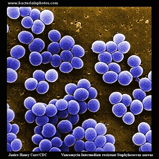
Under a high magnification of 10,000x, this scanning electron micrograph (SEM) shows a strain of Staphylococcus aureus bacteria taken from a vancomycin intermediate resistant culture (VISA,
Vancomycin Intermediate resistant Staphylococcus aureus).
Under SEM, one can not tell the difference between bacteria that are susceptible, or multidrug resistant,
but with transmission electron microscopy (TEM), at least with VISA isolates one can see a thickening in the cell wall
that may attribute to their reduced susceptibility to vancomycin .
Text&Photo CDC, Public Health Image Library (PHIL)
This image is in the public domain and thus free of any copyright restrictions.
Public Health Image Library (PHIL)