| |
Bacteria under Microscope
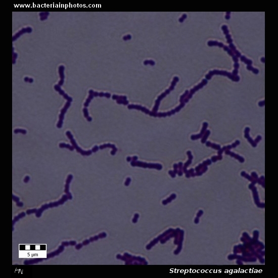
Streptococcus agalactiae
Group B streptococcus, GBS
|
|
|
| |
Gram-stain: |
Gram-positive cocci |
| |
Microscopic appearance: |
Short chains of cocci, diplococci |
| |
Clinical significance: |
- Streptococcus agalactiae is a member of the gastrointestinal normal flora in some humans.
- In the western world, S. agalactiae is the major cause of bacterial septicemia of the newborn, which can lead to death or long-term sequelae.
- S. agalactiae is present in up to one-third of women of childbearing age, and 1.8 cases per 1000 live births will be affected by group B streptococcal infection.
|
| |
|
Text: Wikipedia |
| |
Colony morphology: |
|
|
| |
|
|
|
|
|
A |
B |
C |
|
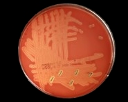
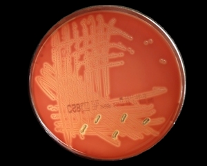
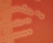
| Streptococcus agalactiae identification |
|
A |
Beta-hemolytic colonies of Streptococcus agalactiae (Group B streptococcus, GBS) on sheep blood agar. Cultivation 24 hours, aerobic atmosphere, 37°C. |
|
|
B |
Colonies of S.agalactiae surrounded by a zone of beta-hemolysis. Colonies of group B streptococci often have less pronounced zones of beta-hemolysis than do other beta-hemolytic streptococci (e.g. from Group A or C); some group B strains are nonhemolytic.
Cultivation 24 hours in an aerobic atmosphere, 37°C. Sheep blood agar |
|
|
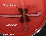
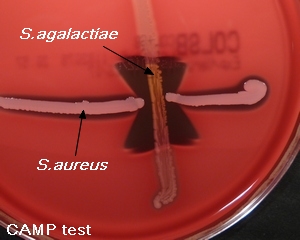
C |
CAMP reaction. Streptococcus agalactiae produce extracellular, diffusible protein (CAMP factor) that acts synergistically with beta-lysin produced by Staphylococcus aureus to produce a zone of enhanced lysis of sheep (or bovine) erythrocytes. Cultivated on Columbia agar with 5% defibrinated sheep blood, 24 hours in an aerobic atmosphere, 37°C.
|
|
|
|
| |
www.bacteriainphotos.com |
|
|