CULTIVATION MEDIA IN BACTERIOLOGY I |
| |
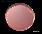
MacConkey agar |
|
|
MacConkey agar plate
|
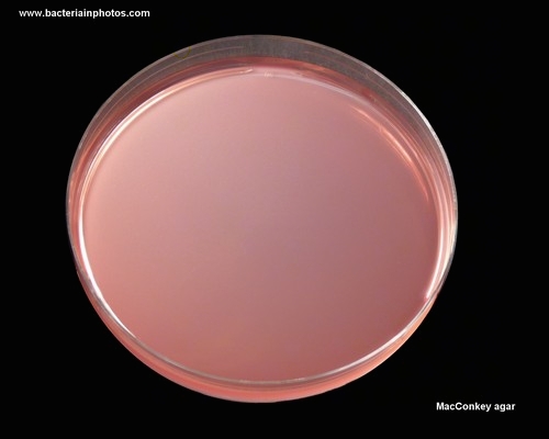
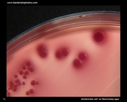
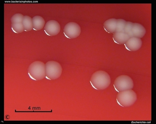
Escherichia coli
on MacConkey agar
(lactose positive)
|
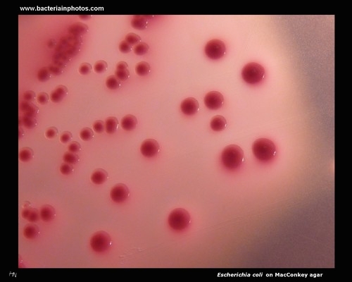
Lactose fermenting
Escherichia coli
on MacConkey agar
|
|
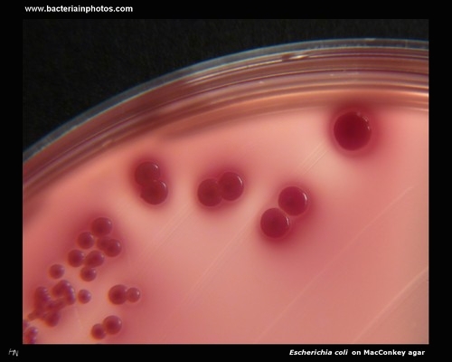
Appearance of lactose positive E.coli on MacConkey
|
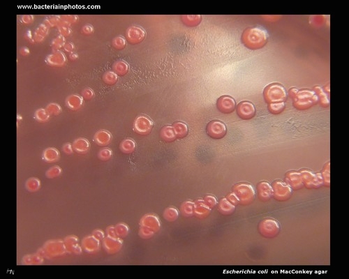
MacConkey agar: lactose positive colonies
of Escherichia coli
|
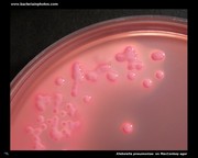
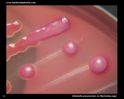
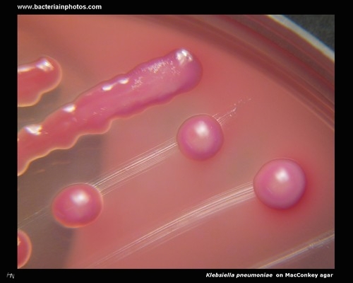
Klebsiella pneumoniae
on MacConkey:
lactose positive
|
|
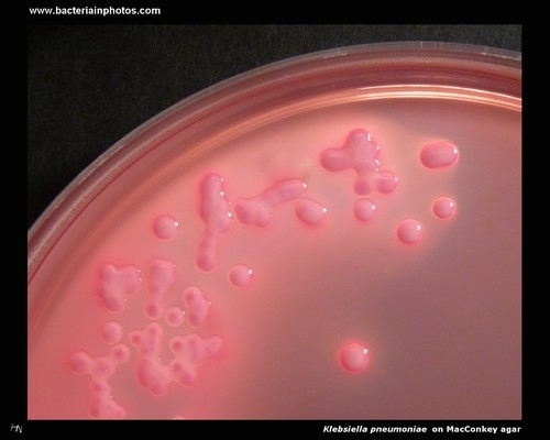
Colonies of
Klebsiella pneumoniae on MacConkey agar
(lactose positive)
|
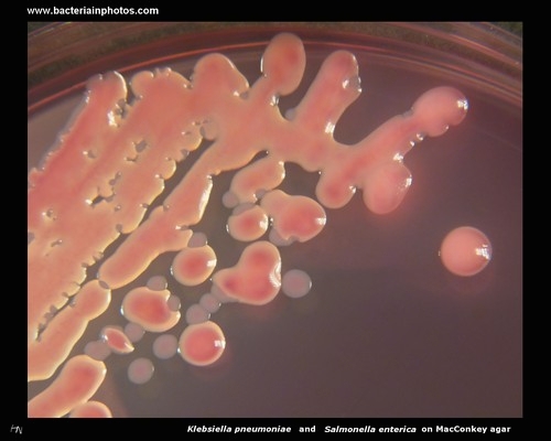
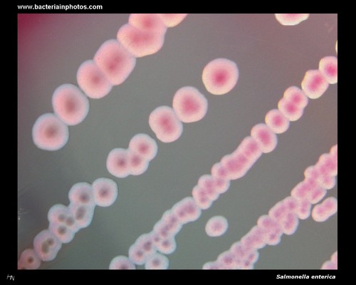
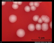
Klebsiella pneumoniae and Salmonella enterica
on MacConkey agar:
lactose + and -
|
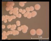
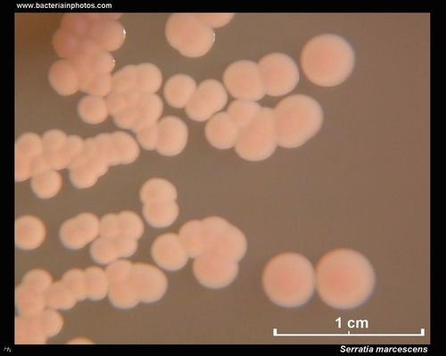
Serratia marcescens
on MacConkey agar:
lactose negative
|
Lactose negative Pseudomonas aeruginosa
on MacConkey agar
|
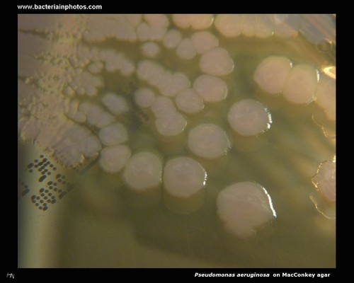
Pseudomonas aeruginosa
on MacConkey agar
(lactose negative)
|
MacConkey Agar
Use: Moderately selective culture medium for the detection of coliform organisms and enteric pathogens
|
| GROWTH |
INHIBITION OF GROWTH |
LACTOSE POSITIVE
examples |
LACTOSE NEGATIVE
examples |
|
- Escherichia coli (most strains)
- Klebsiella pneumoniae
- Entrobacter cloacae
|
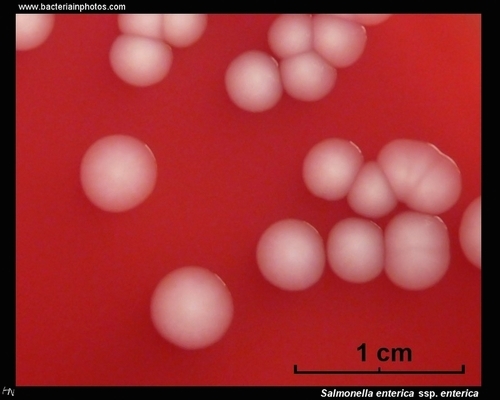
- Salmonella enterica ssp. enterica
- Shigella spp.
- Proteus spp.
- Citrobacter freundii (some strains)
- Morganella morganii
- Providencia spp.
|
- staphylococci
- streptococci
- enterococci
Crystal violet and bile salts in medium inhibit growth of Gram-positive microorganisms
|
| BACTERIUM |
TYPICAL GROWTH ON MACCONKEY AGAR |
| Escherichia coli |
Pink to rose-red. Colonies may be surrounded by a zone of precipitated bile. |
| Enterobacter cloacae |
Pink, mucoid. |
| Klebsilla pneumoniae |
Pink, mucoid. |
| Proteus |
Colorless. |
| Pseudomonas aeruginosa |
Colorless to pink. |
| Salmonella and Shigella |
Colorless. |
| Gram-positive bacteria |
No growth to slight growth (pale pink). |
|
|
| MacConkey agar on Wikipedia |
| www.microbelibrary.org |
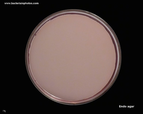
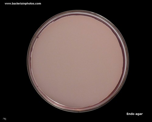
Endo Agar |
|
Endo agar plate
|
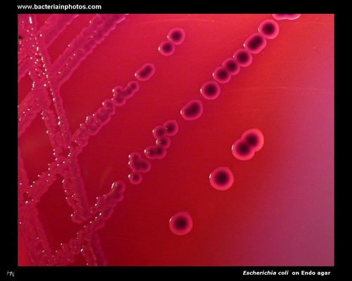
Lactose positive colonies
of Escherichia coli
on Endo agar
|
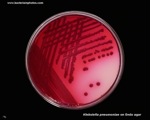
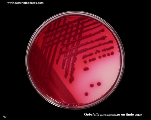
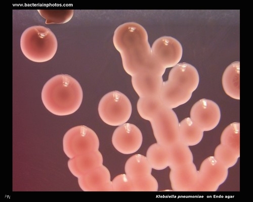
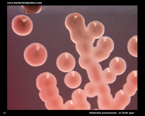
Lactose positive colonies
of Klebsiella pneumoniae
on Endo agar
|
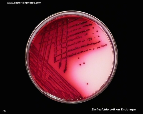
Colonies of Escherichia coli
on Endo agar
(lactose positive)
|
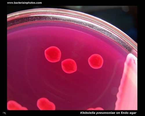
Colonies of Klebsiella pneumoniae
on Endo agar
(lactose positive)
|
|
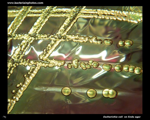
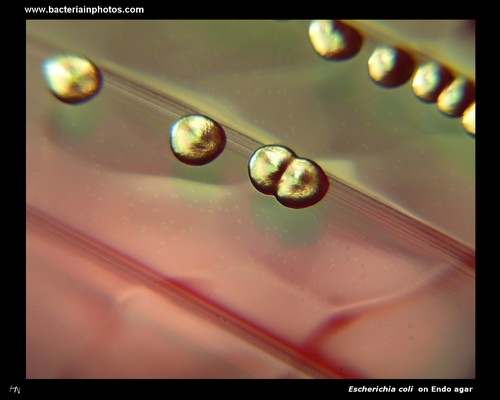
Colonies of
Escherichia coli on Endo agar
(lactose positive, metallic sheen)
|
E.coli on Endo agar
(lactose positive, metallic sheen)
|
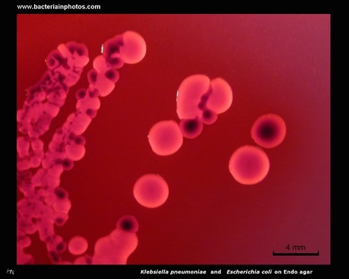
Klebsiella pneumoniae and Escherichia coli on Endo agar (lactose positive)
|
Klebsiella pneumoniae
on MacConkey agar (lactose positive)
|
Lactose negative colonies
of Salmonella enterica
on Endo agar
|
|
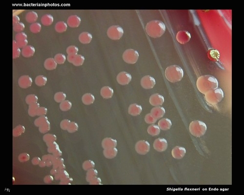
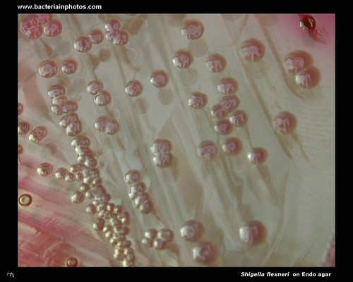
Shigella flexneri on Endo agar
(lactose negative)
|
Colonies of
Shigella flexneri on Endo agar
(lactose negative) |
|
|
|
Endo Agar
Use: Moderately selective culture medium for the detection and confirmation of coliforms and other enteric microorganisms
|
| GROWTH |
INHIBITION OF GROWTH |
LACTOSE POSITIVE
examples |
LACTOSE NEGATIVE
examples |
|
- Escherichia coli (most strains)
- Klebsiella pneumoniae
- Entrobacter cloacae
|
- Salmonella enterica ssp. enterica
- Shigella spp.
- Proteus spp.
- Citrobacter freundii (some strains)
- Morganella morganii
- Providencia spp.
|
- staphylococci
- streptococci
- enterococci
Inhibition of Gram-positive microorganisms is achieved by the sodium sulfite and basic fuchsin contained in the formulation
|
| BACTERIUM |
TYPICAL GROWTH ON ENDO AGAR |
| Escherichia coli |
Colonies pink to rose-red with/without green metallic sheen. |
| Enterobacter cloacae |
Pink to rose-red, mucoid. |
| Klebsilla pneumoniae |
Pink to rose-red, mucoid. |
| Proteus |
Colorless. |
| Pseudomonas aeruginosa |
Colorless. |
| Salmonella and Shigella |
Colorless. |
| Gram-positive bacteria |
No growth to slight growth (pale pink to red). |
|
|
| Endo agar on Wikipedia |
Deoxycholate Citrate Agar |
|
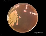
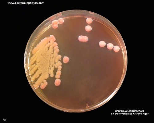
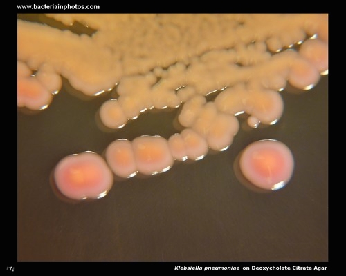
Klebsiella pneumoniae
on Deoxycholate Citrate Agar (DCA)
(lactose positive)
|
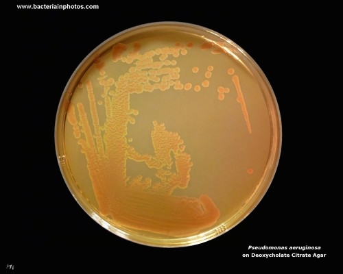
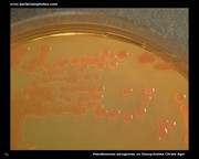
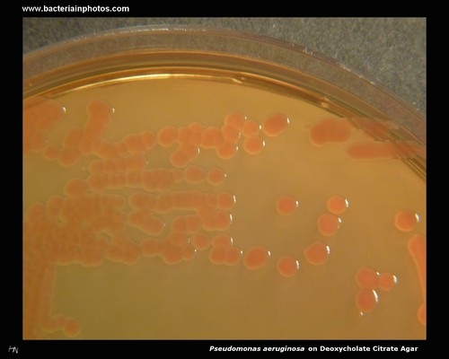
Pseudomonas aeruginosa
on Deoxycholate Citrate Agar
(lactose negative)
|
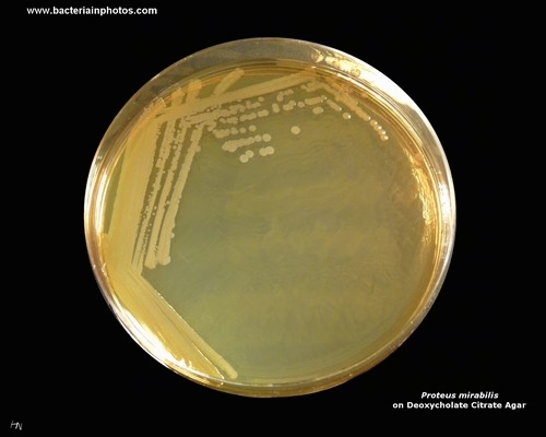
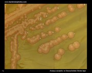
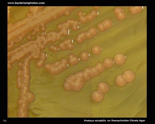
Proteus mirabilis on Deoxycholate Citrate Agar
(lactose negative)
|
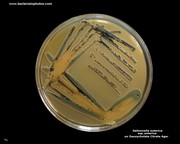
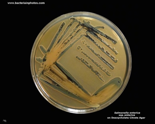
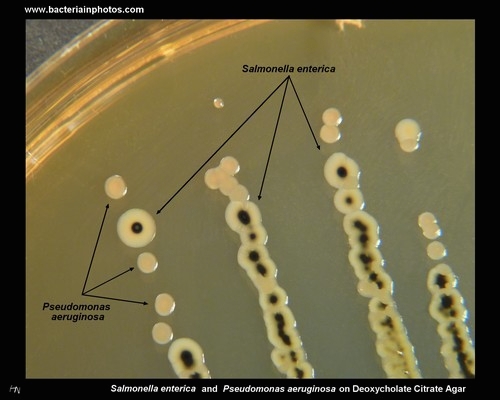
Salmonella enterica
on Deoxycholate Citrate Agar
(lactose negative,
H2S positive)
|
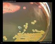
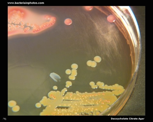
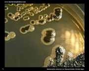
Lactose negative and lactose positive colonies of bacteria on Deoxycholate Citrate Agar (DCA)
|
|
Klebsiella pneumoniae
on Deoxycholate Citrate Agar
(lactose positive)
|
Proteus mirabilis
on Deoxycholate Citrate Agar
(lactose negative)
|
Pseudomonas aeruginosa
on Deoxycholate Citrate Agar
(lactose negative)
|
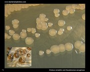
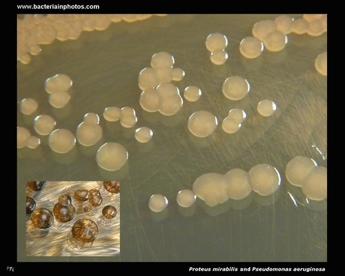
Lactose negative colonies of Proteus mirabilis (larger)
and Pseudomonas aeruginosa on Deoxycholate Citrate Agar
|
Lactose negative, H2S positive colonies of Salmonella enterica on Deoxycholate Citrate Agar
|
|
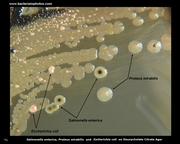
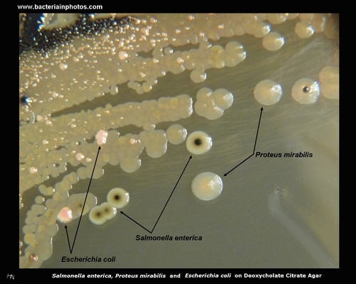
Lactose negative, H2S positive colonies of Salmonella enterica, lactose negative colonies of Proteus mirabilis and lactose positive colonies of Escherichia coli
|
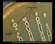
Lactose negative, hydrogen sulfide positive colonies of Salmonella enterica and lactose negative colonies of Pseudomonas aeruginosa
|
|
|
|
Deoxycholate Citrate Agar
Use: Isolation of Salmonella and Shigella species; moderately selective.
|
| GROWTH |
INHIBITION OF GROWTH |
LACTOSE POSITIVE
examples |
LACTOSE NEGATIVE
examples |
|
- Escherichia coli (most strains)
- Klebsiella pneumoniae
- Entrobacter cloacae
Partial to complete inhibition.
|
- Salmonella enterica ssp. enterica
- Shigella spp.
- Proteus spp.
- Pseudomonas aeruginosa
|
Completely suppress the Gram-positive microbial flora.
Inhibition of Gram-positive microorganisms and coliforms is achieved by the deoxycholate and citrate contained in the formulation.
|
| BACTERIUM |
TYPICAL GROWTH ON DEOXYCHOLATE CITRATE AGAR |
| Salmonella spp. |
Colorless colonies with or without black centers (H2S production). |
| Shigella spp. |
Colorless. |
| Proteus |
Colorless colonies with or without black centers (H2S production). Partial to complete inhibition. |
| Escherichia coli |
Pink with/without bile precipitate. |
| Enterobacter cloacae |
Pink. Partial to complete inhibition. |
| Klebsiella pneumoniae |
Pink. Partial to complete inhibition. |
| Pseudomonas aeruginosa |
Colorless. Partial to complete inhibition. |
| Gram-positive bacteria |
No growth. |
|
|
| Deoxycholate Citrate Agar on Wikipedia |
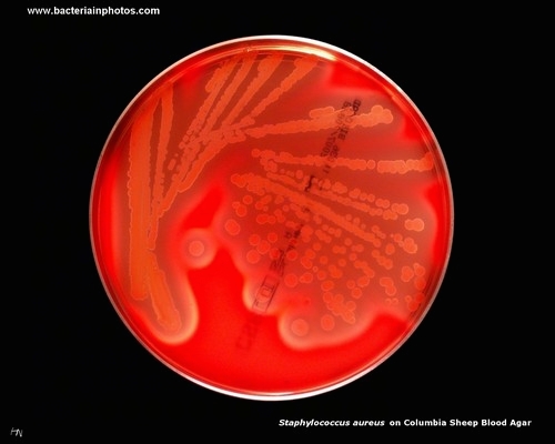
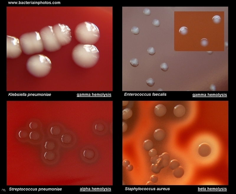
Blood Agar |
|
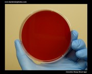
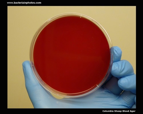
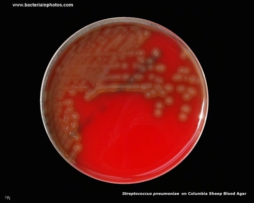
Columbia Sheep Blood Agar
|
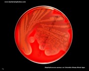
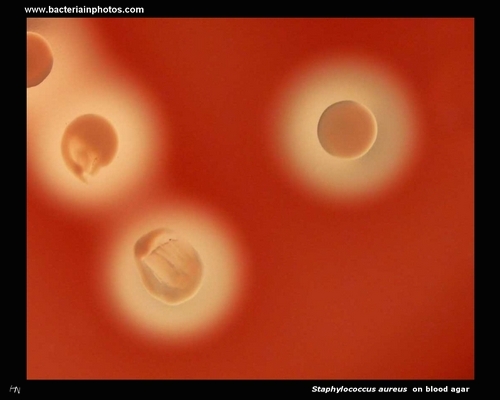
Beta-hemolysis on blood agar (Staphylococcus aureus)
|
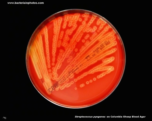
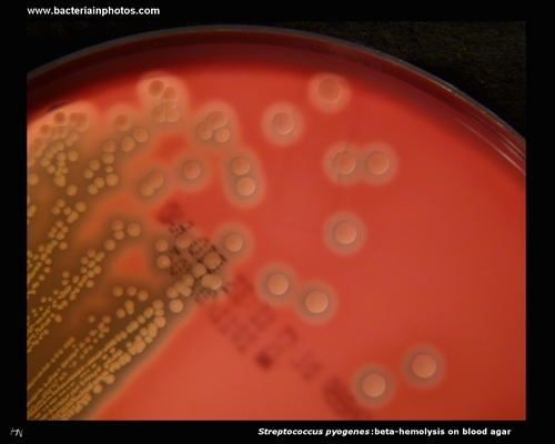
Beta-hemolysis on blood agar (Streptococcus pyogenes)
|
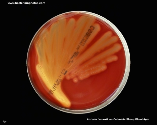
Beta-hemolysis on blood agar (Listeria ivanovii)
|
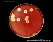
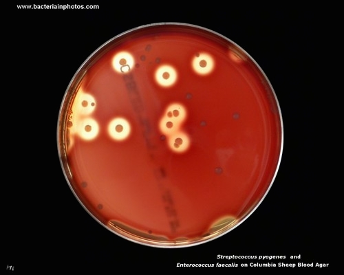
Beta-hemolysis (Streptococcus pyogenes) and gamma-hemolysis (Enterococcus faecalis) on blood agar
|
|
|
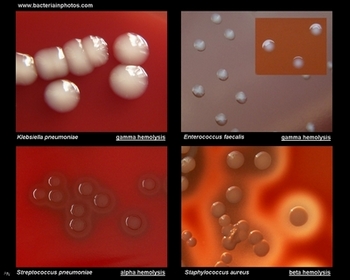
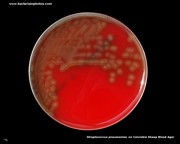
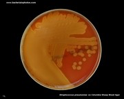
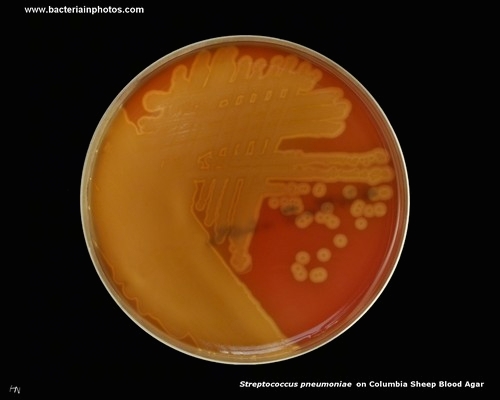
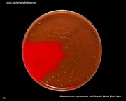
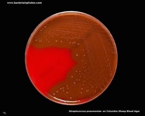
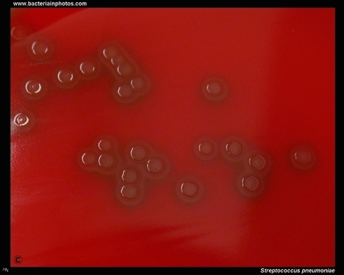
Alpha-hemolysis on blood agar (Streptococcus pneumoniae)
|
Alpha-hemolysis on blood agar (Streptococcus pneumoniae)
|
Alpha-hemolysis on blood agar (Streptococcus pneumoniae)
|
|
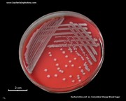
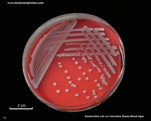
Gamma-hemolysis on blood agar (Escherichia coli)
|
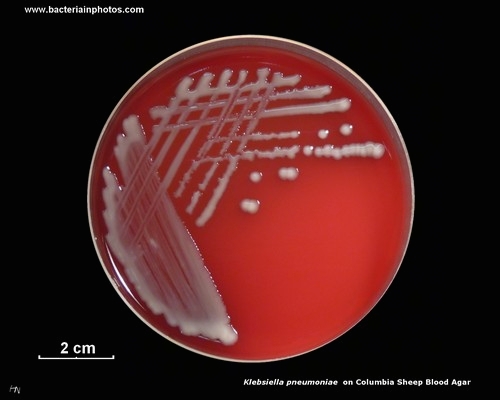
Gamma-hemolysis on blood agar (Klebsiella pneumoniae)
|
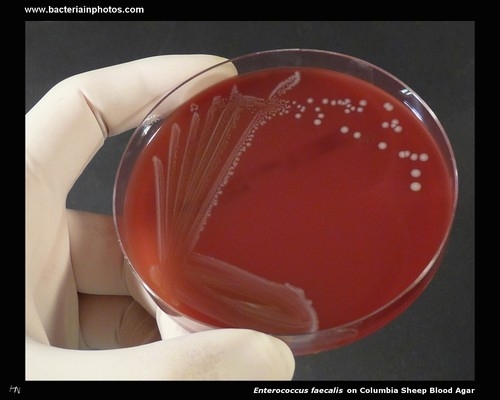
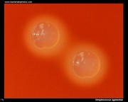
Gamma-hemolysis on blood agar (Enterococcus faecalis)
|
|
Non-hemolytic colonies on blood agar
(Salmonella enterica)
|
Gamma-hemolysis on blood agar (Escherichia coli)
|
Alpha-hemolysis on blood agar (Streptococcus pneumoniae)
|
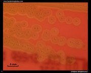
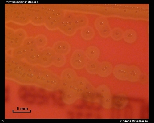
Alpha-hemolysis on blood agar (viridans streptococci)
|
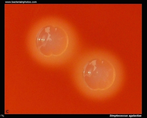
Beta-hemolysis on blood agar (Streptococcus agalactiae)
|
|
Beta-hemolytic colonies on blood agar
(Staphylococcus aureus)
|
Beta-hemolysis on blood agar (Streptococcus pyogenes)
|
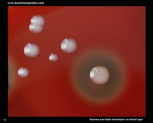
Gamma-hemolysis and beta-hemolysis on blood agar (Enterococcus faecalis and Streptococcus pyogenes)
|
|
|
| Blood agar on Wikipedia |
| Hemolysis on Wikipedia |
| www.microbelibrary.org |
Mueller Hinton Agar |
|
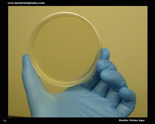
Mueller Hinton agar
|
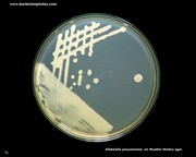
Klebsiella pneumoniae
on Mueller Hinton agar
|
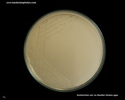
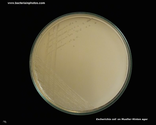
Escherichia coli
on Mueller Hinton agar
|
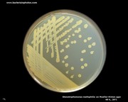
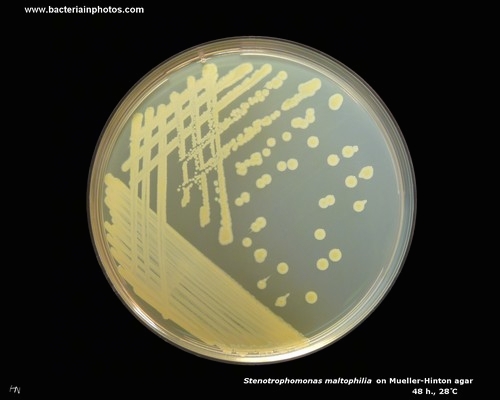
Stenotrophomonas maltophilia
on Mueller Hinton agar
|
Staphylococcus aureus
on Mueller Hinton agar
|
|
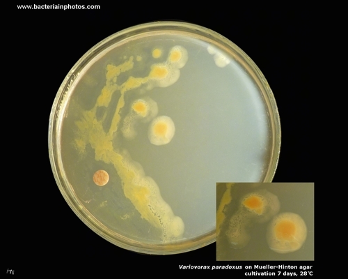
Variovorax paradoxus
on Mueller Hinton agar
|
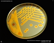
Chryseobacterium indologenes
on Mueller Hinton agar
|
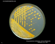
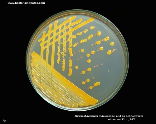
Chryseobacterium indologenes
and an actinomycete
on Mueller Hinton agar
|
|
|
Yellowish colonies of
Stenotrophomonas maltophilia
on Mueller Hinton agar
|
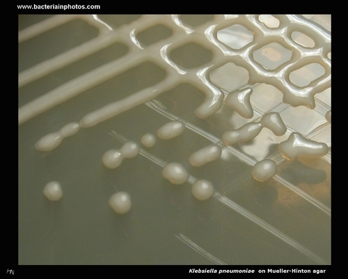
Mucoid colonies of
Klebsiella pneumoniae
on Mueller Hinton agar
|
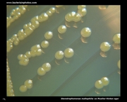
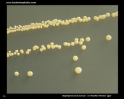
Yellow colonies of
Staphylococcus aureus
on Mueller Hinton agar
|
|
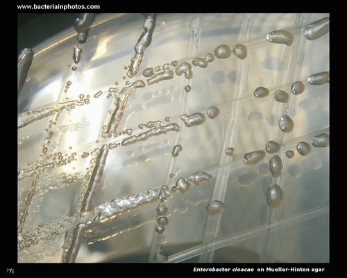
Enterobacter cloacae
on Mueller Hinton agar
|
Rapidly growing
Mycobacterium sp.
on Mueller Hinton agar
|
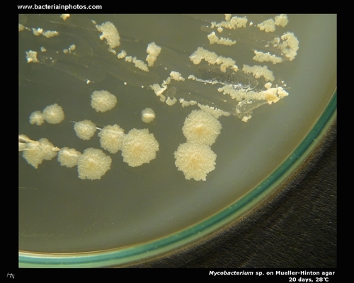
Colonies of Mycobacterium sp.
on Mueller Hinton agar
(eugonic growth)
|
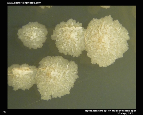
Mycobacterium sp.
on Mueller-Hinton agar
|
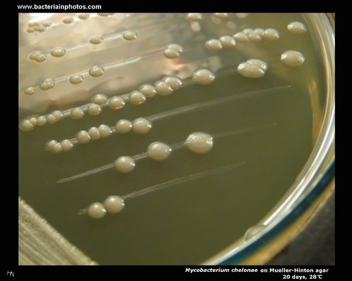
Mycobacterium chelonae
on Mueller Hinton agar
(disgonic growth)
|
| Mueller Hinton agar on Wikipedia |
Disk Diffusion Test on Mueller Hinton Agar |
|
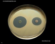
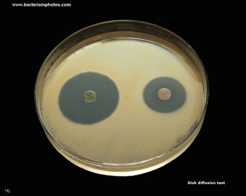
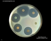
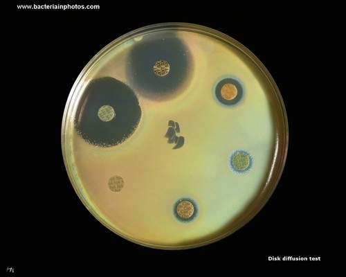
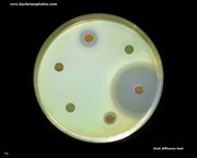
Disk Diffusion Test
on Mueller-Hinton Agar
|
Bacterium susceptible
to ampicillin and resistant to gentamicin
|
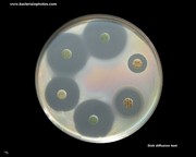
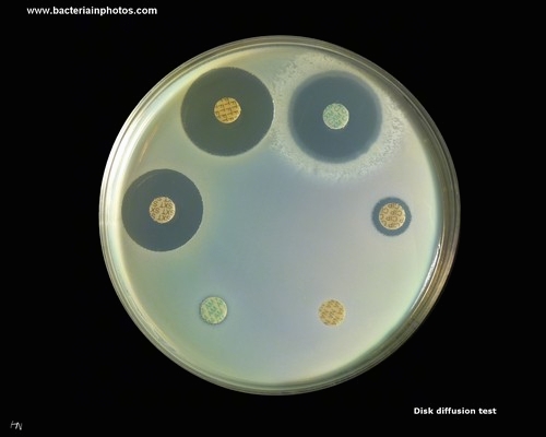
Good susceptibility to antibiotics
|
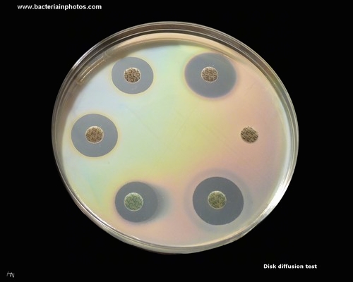
Kirby-Bauer antibiotic testing
on Mueller-Hinton Agar
|
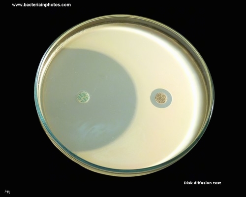
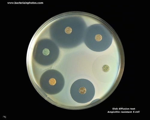
Disk Diffusion Test
on Mueller-Hinton Agar
(susceptible strain, ampicillin resistant)
|
|
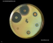
Bacterium resistant to 3 out of 6 tested antibiotics
|
Alcaligenes faecalis
resistant to 4 out of 6 tested antibiotics
|
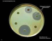
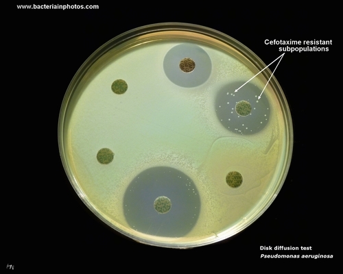
Pseudomonas aeruginosa:
subpopulations of cefotaxime
resistant bacteria
|
Pseudomonas aeruginosa:
susceptible to ciprofloxacin,
resistant to the rest
(small inhibition zone around tetracycline)
|
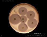
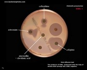
Klebsiella pneumoniae:
good susceptibility
to tested antibiotics
(ESBL screening)
|
|
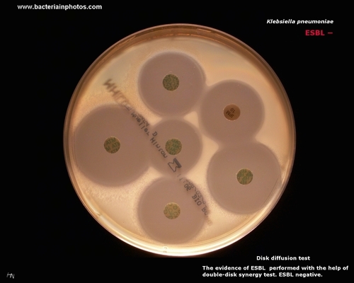
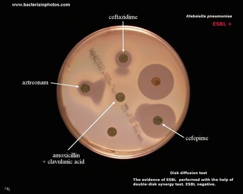
Klebsiella pneumoniae:
double disk synergy test
(ESBL positive)
|
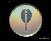
Staphylococcus aureus:
E-test
|
|
| Antibiotic testing (on Wikipedia) |
| E-test(on Wikipedia) |
| www.microbelibrary.org |